
Virus Destroying Plastic, Helmets as hard as Nuts and Cancer Detection from Blood Samples
May 7
This week we discover a new this plastic covering that can tear apart viruses upon impact. We examine how a Western Australian native nut inspired a new type of material for the next generation of protective gear. We learn about a new paint that changes color depending on how hard it is hit. Finally we investigate a new blood test that can detect many different types of cancer and other conditions.
Plastic Film that Destroys Viruses
A team at RMIT in Melbourne have developed a plastic surface with tiny nanoscale features billionths of a meter in size that can physically rupture viruses. The design mimics the nano textured surface of insect wings. The material offers a cheap scalable way to make surfaces such as phones and hospital equipment far less likely to spread disease.
We currently combat viruses by cleaning to remove dirt and disinfection to kill hidden contaminants. Disinfectant must remain wet for some time to kill germs. Additionally the surface only remains clean until the next person touches it. At best disinfectant is a short term solution.
Other attempts at antiviral surfaces have incorporated materials such as graphene or tannic acid into personal protective equipment such as masks, gloves, goggles, hard hats and respirators. Whilst efficient these coatings can pose a risk to human health. Chemical leaching harms the environment and these materials lose effectiveness over time as the potency of the active ingredients weakens.
Nature has examples of bacteria free surfaces. The water repelling wings of cicadas and dragonflies are self cleaning and kill bacteria. It is the physical nanostructure on the surface that forces the bacterial cell membranes to stretch and rupture. The team had previously developed a nano spike covered silicon that destroys viruses on contact however the rigid nature restricts its use on complex objects.
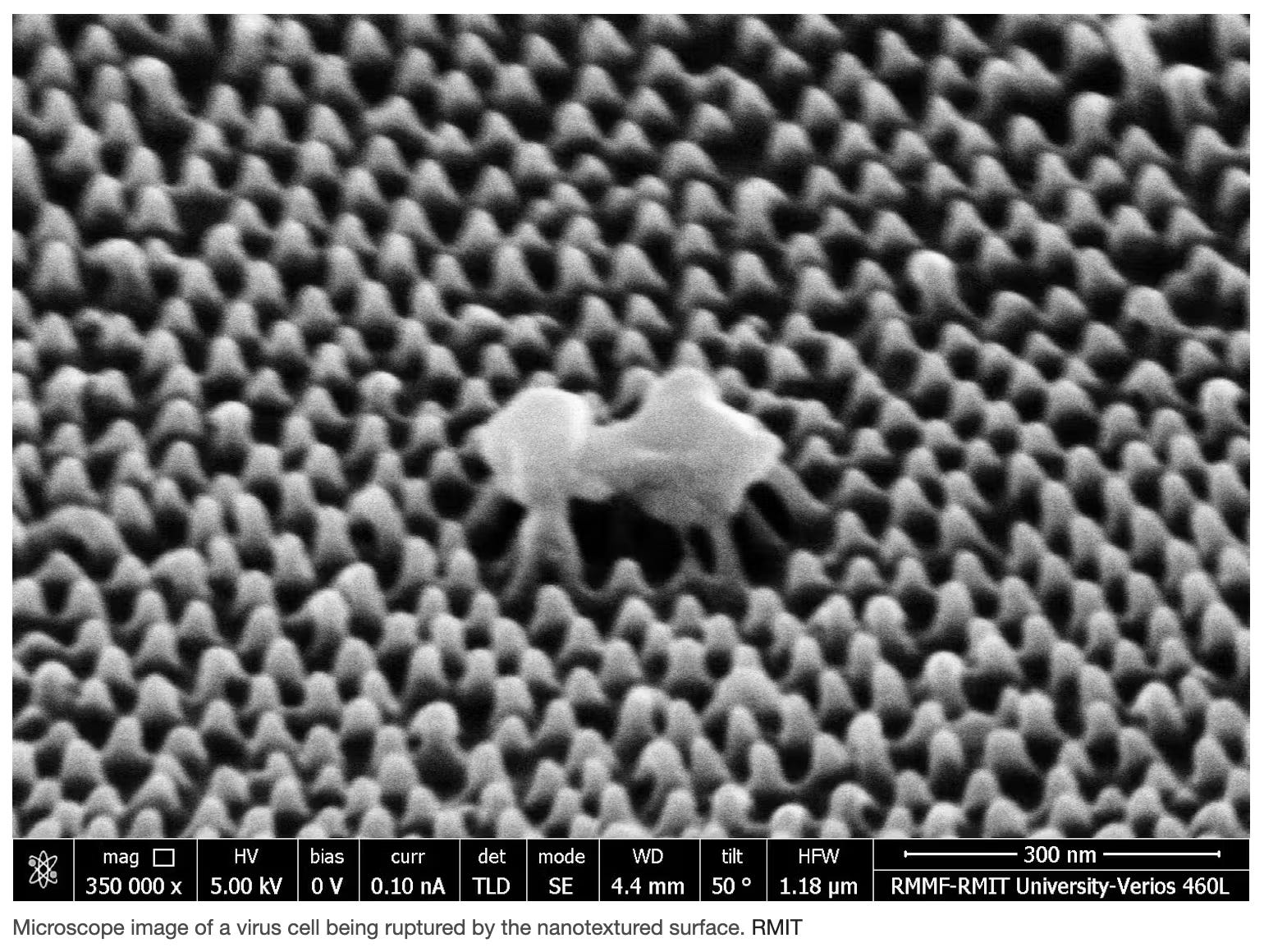
The team set out to create a virus busting material that was lightweight, cost effective and flexible. The result was a thin acrylic film covered in thousands and thousands of ultra fine pillars. The pillars grab and stretch a virus’s outer shell until it ruptures. The film kills the virus through mechanical force.
Lab tests with a human flu virus showed 94% of virus particles were ripped apart or fatally damaged within an hour of contact with the material. The team found that the distance between pillars was more important than the hight. The film feels smooth to human touch.
The film was created using a mold that can be easily scaled for efficient manufacturing. Potential uses include hospitals, public transport systems, office desks and food packaging.
Helmets like Nuts
A team at NYU Abu Dhabi has uncovered the secret to the remarkable toughness of the marri nut, a tree native to Western Australia. The shell is so strong that predators snuggle to break it. The team studied how the nut absorbs impact and resists cracking. The secret is in a cleaver natural design that may provide inspiration for the next generation of protective gear.
The nut’s strength comes from a layered internal structure that combines a tough outer shell with a softer flexible interior. The combination of hardness and flexibility allows the nut to absorb energy without shattering. Force is distributed in a controlled way rather than breaking suddenly. Cracks are not allowed to spread.
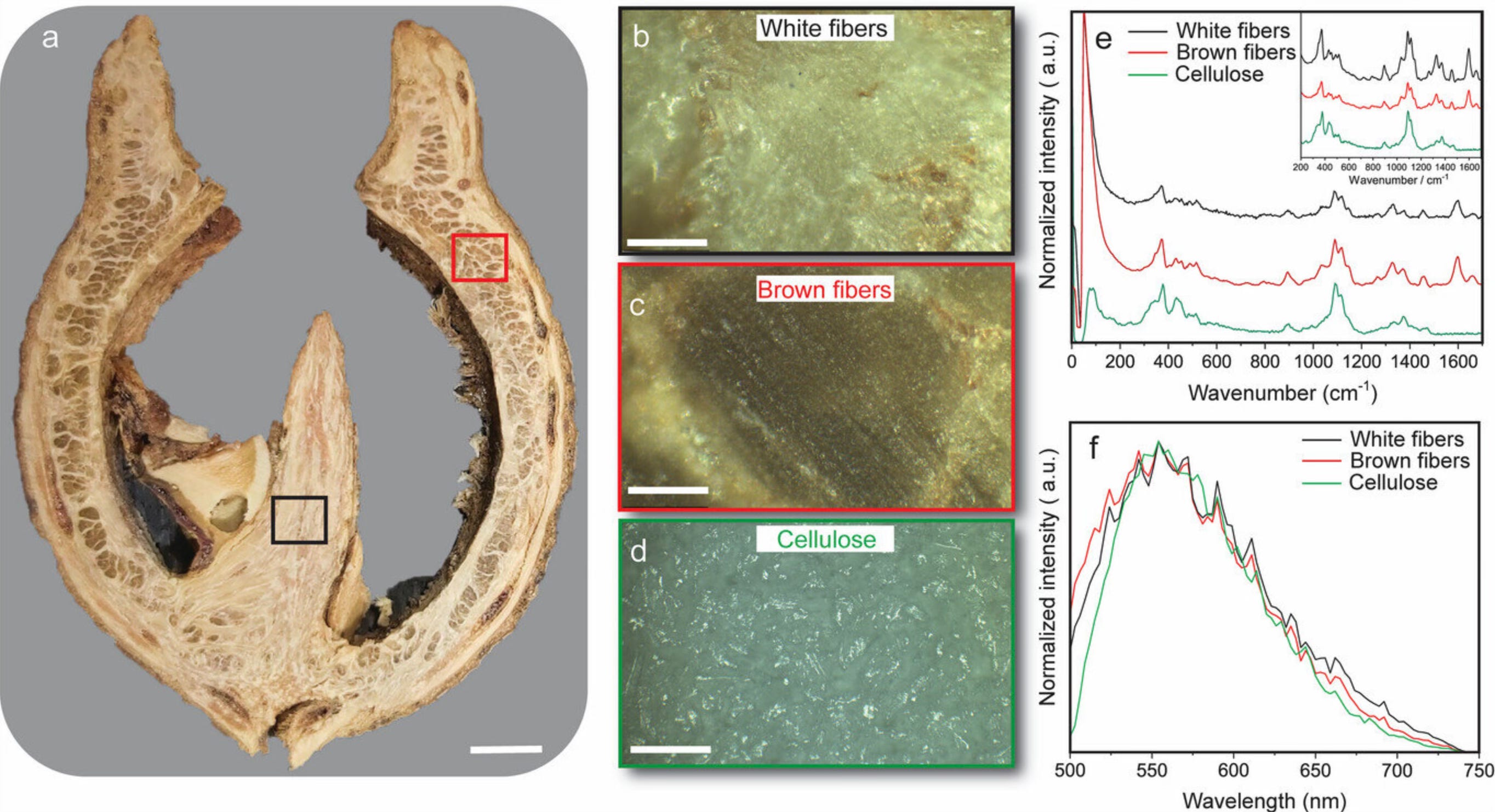
The natural cellulose material can deform and absorb energy like Teflon whilst maintaining stiffness similar to acrylic whilst remaining lightweight. The researchers were able to create a material that mimics the nut’s internal structure. A new generation of safety equipment may soon be upon us. Another example of nature inspiring science.
Early Detection of Cancer from Blood Samples
A team at UCLA have developed a simple cost effective blood test that in early trials has shown promise in detecting multiple cancers, various liver conditions and organ abnormalities. The test analyzes DNA fragments circulating in the blood stream.
Called MethyScan the test works by analyzing cell free DNA. These are tiny fragments of genetic material released into the blood when cells die. Cells from every organ shed DNA into the bloodstream. This cell free DNA carries molecular signals that reflect what is happening throughout the body.
Every day 50 to 70 billion cells in our bodies die. These cell’s DNA goes into the bloodstream providing information from all our organs via our blood. There are some tests that look for mutations in tumor DNA to screen for certain cancers. These tests focus on a limited number of genetic changes and are expensive due to the requirement to sequence DNA to detect faint tumor signals.
Rather than search for mutations, the team examined DNA methylation chemical tags attached to DNA that help regulate gene activity. Methylation patterns differ by tissue type and can change when cells become cancerous or diseased.
One challenge that the team had to overcome was the 80% to 90% of cell free DNA originates from normal blood cells not tumors or injured organs. A technique was developed to remove much of this blood cell background DNA before sequencing. Using specialized enzymes they selectively cut away un-methylated DNA fragments. The result is that the captured DNA fragments come mostly from solid organs.
The resulting sequencing only required 5GB of data which would cost US$20 at current rates (this will only fall). In a test of 1061 patients with liver, lung, hepatitis C, alcohol related liver disease and people with benign lung nodules and healthy participants the test detected 63% of cancers across all stages with 55% of early stage cancers found. A high specificity rate of 98% reduced the number of false positives significantly.
The ability to trace signals back to the source organ allows focussed follow up. Larger trials will be needed to confirm the test’s performance in real world conditions.
Paint that Changes Color when Hit
A team at Tufts University in Massachusetts have developed a paint that changes color depending upon how hard its surface has been hit. It could be used in American Football helmets to monitor concussion potential, to record the handling of packages that are delivered and to analyze our gait by painting the soles of our shoes.
The paint was developed to quantitatively measure the site and force of an impact without using any electronic circuitry or sensors. A color changing polymer surrounded by a silk protein polymer shell can be painted on any surface of almost any size, texture or contour.
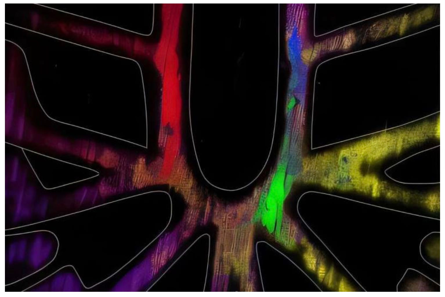
The paint contains tiny spherical particles each the size of a human blood cell. The particles contain a core of color changing polymer surrounded by a harder polymer shell made of silk fibroin proteins derived from the common silk moth. The core polymer undergoes a blue to red transition when under mechanical stress. The stress twists the chemical backbone of the inner polymer affecting how electrons move along its length. This affects how photons are absorbed which changes the colors of light absorbed and emitted.
The amount of red released increases with how hard the surface of the paint particles are hit. This allows the paint to act as a built in force meter. The hardness of the outer shell can be tuned to extend the response of the paint to different levels of force. Once the color changes it stays changed. The level of the color change can be converted directly into newtons, a unit of force.
Paying it Forward
If you have a start-up or know of a start-up that has a product ready for market please let me know. I would be happy to have a look and feature the startup in this newsletter. Also if any startups need introductions please get in touch and I will help where I can.
If you have any questions or comments please comment below.
I would also appreciate it if you could forward this newsletter to anyone that you think might be interested or provide a recommendation on Substack.